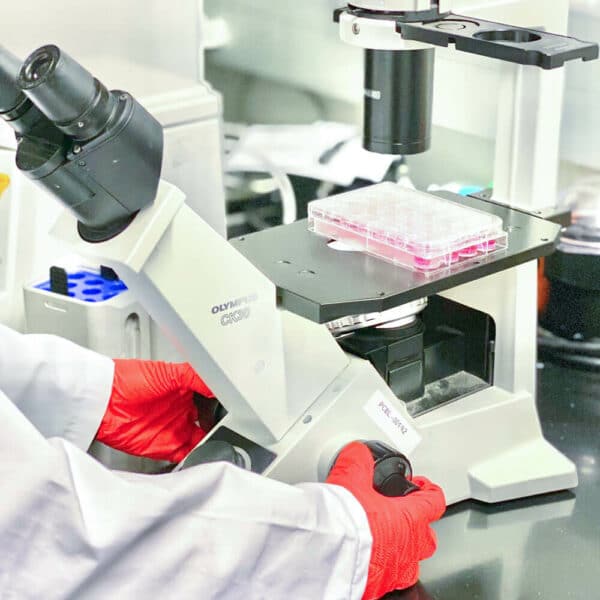

“3D Bioprinting” or “bioprinting” is a form of additive manufacturing that uses cells and biomaterials instead of traditional metals and plastics to create 3D constructs that are functional 3D tissues. These biomaterials are called bioinks, and they mimic the composition of our tissues. Bioprinting can be applied to a variety of areas including but not limited to regenerative medicine, drug discovery and development, and 3D cell culture.
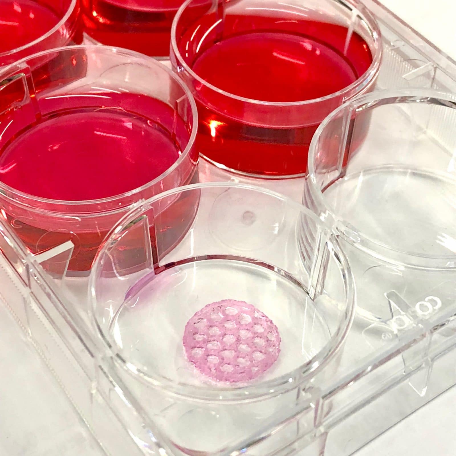
Bioprinted structures, such as organs-on-chips, can be used to study functions of a human body outside the body, in 3D. The geometry of a 3D bioprinted structure is more similar to that of a naturally occurring biological system than an in vitro 2D model. Structural similarity can in turn lead to functional results that are more physiologically relevant. No other technology enables the level of geometric complexity in engineering tissues that 3D bioprinting enables. That is why this technology has the potential to completely change the way we treat diseases – by replacing animal testing and ending the organ transplant waiting list.
How does 3D bioprinting work?
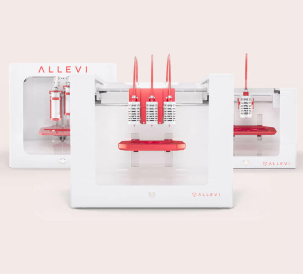
3D bioprinting starts with a model of a structure, which is recreated layer-by-layer out of a bioink either mixed with living cells, or seeded with cells after the print is complete. These starting models can come from anywhere – a CT or MRI scan, a computer generated design (CAD) program, or a file downloaded from the internet.
That 3D model file is then fed into a slicer – a specialized kind of computer program which analyzes the geometry of the model and generates a series of thin layers, or slices, which form the shape of the original model when stacked vertically. Cura and slic3r are examples of slicers commonly used in 3D printing. Allevi also has a specialized slicer, optimized specifically for bioprinting, built into our Allevi Bioprint software.
Once a model is sliced, the slices are transformed into path data, stored as a G-code file, which can be sent to a 3D bioprinter for printing. The bioprinter follows instructions in the G-code file in order, including instructions to control for temperature of the extruders, extrusion pressure, bed plate temperature, crosslinking intensity and frequency, and, of course, the 3D movement path generated by the slicer. Once all of the G-code commands are completed, the print is done and can be cultured or seeded with cells as part of a biostudy.
Why is bioprinting important?
Over 120,000 people in the US alone are on waiting lists for organs, and others experience chronic problems due to the long-term damaging effects of post-transplant immunosuppression. There is a large and growing need for an alternative to the organ transplant waiting list. The scientific community has already succeeded in bringing together multidisciplinary teams of researchers, physicians, and engineers to take on the biggest challenges to human health, and 3D bioprinting is an exciting new tool with the potential to eliminate the organ transplant waiting list.
For pharmaceutical development, 3D bioprinting offers a means of testing drugs faster, at a lower cost, and with better biological relevance to humans than animal testing. In the biomedical devices field, 3D bioprinting has enabled new developments such as sugar stents to help surgeons join veins with fewer complications, and systems for improved drug delivery, among others.
As bioprinting evolves, it will become possible to use a patient’s own cells to 3D print skin and bone grafts, organ patches, and even full replacement organs. Personalized and regenerative medicine continue to grow in popularity, and 3D bioprinting will give doctors and researchers the tools to better target treatments and improve patient outcomes.
Further reading
Bioprinting 101 – Allevi
3D Bioprinting – Wikipedia
3D Bioprinting of Living Tissues – Wyss Institute, Harvard
Bioprinting – NIH Director’s Blog